The abdominal cerci of orthoptera bear mechanoreceptive hairs that allow the insect to perceive air currents generated by an attacking predator. Crickets own the most elaborated cercal system among orthopthera. I focus my study on a particular species Nemobius sylvestris living in the litter of forest. This cricket can perceive the air movements generated by his natural predator the wolf spider Licosidae Pardosa sp. in noisy environment (Figure 1). I study at the behavioural and neurobiological level its perception capabilities, both in controlled conditions in the laboratory and in its noisy natural environment the forest. I used a controlled piston to stimulate the cercal system (Figure 2). Using behavioural experiments I characterized the escape behaviour and I studied implication of different sensory organs like cerci, antennae, vision. The neurobiological studies are done using extra-cellular electrophysiological
Publication List:
Dupuy F., Josens R., Giurfa M. And Sandoz J.C. (2010) Calcium imaging in the ant Camponotus fellah reveals a conserved odour-similarity space in insects and mammals. BMC Neuroscience 11-28.
Dupuy F., Casas J., Bagnères A.G., Lazzari C.R., (2009), OpenFluo: A free open-source software for optophysiological data analyses, Journal of Neuroscience Methods, 183:195-201.
Dupuy F., Sandoz J.C., Giurfa M. and Josens R.. (2006) Individual olfactory learning in Camponotus ants.
Animal Behaviour 72:1081-1091.
Freeware:
Dr. Fabienne Dupuy

2009-to present. Post-doc, Department of zoology, University of Cambridge (UK).
2005-2009. PhD student, IRBI (France).
2003-2004. Master in Neurosciences, Université Paul Sabatier,
Keywords:
Sensory systems; Neurosciences; Insect behaviour; Nervous
Behavioural and neurobiological study of the wood cricket’s mechanosensory system


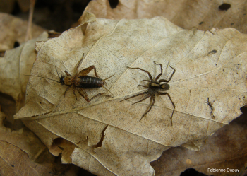
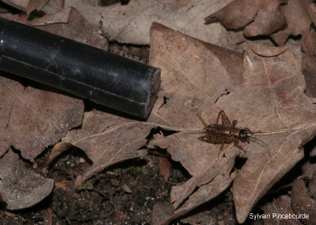

Figure 1. The wood cricket is preyed upon by the wolf spider at the litter surface.
Figure 2. The piston is used to simulate the spider attack in terms of air-flow.
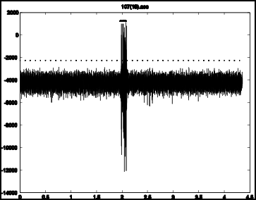
Figure 3. Electrophysiological recordings: [left] The electrode (here the shiny tiny cable) is attached to the connective which leaves the terminal abdominal ganglion.. [right] Spikes of neuronal activity were recorded as the piston approached the cricket.